When you visit your optometrist for an eye exam, you can expect a series of different tests. These allow your optometrist to fully understand your eye, how it works, whether you have any problems, and whether you’re showcasing any signs of potential eye conditions. One such test is called “retinal imaging,” but why is it an important part of an eye exam?
Retinal imaging allows your optometrist to thoroughly examine the light-sensitive tissue at the back of your eye. This tissue can be affected by several conditions, like age-related macular degeneration, diabetes, and more.
By examining this tissue, your optometrist is working towards fully understanding your eye and can address potential problems long before they permanently affect your vision.
What Is the Retina?
Your eye is a wonderfully complex part of the body. It lets light enter through the clear front lens. This light is refracted to reach a focal point in the back of your eye on the light-sensitive tissue called the “retina.” The retina then converts the light into signals that can be sent through the optic nerve to the brain. This is the basics of your visual system.
The retina has 2 primary types of cells that help to process this light. These are called “rods” and “cones.” The rods allow you to see in low-light conditions and help give you your peripheral vision. Meanwhile, the cones in the macula are responsible for helping you see colour. When working properly, the retina is responsible for a significant part of your visual system.
However, these signals can’t be sent properly if the retina is damaged or affected. Instead of a crisp, clear image that accurately portrays your surroundings, this image could be blurry, cloudy, dark, or distorted. This makes it almost impossible to see the world around you clearly.
Why Is the Retina Important?
The importance of the retina cannot be overstated—it plays a critical role in vision. But it’s also easily affected by a range of systemic and ocular conditions, such as:
- Diabetes: This can lead to diabetic retinopathy, a condition in which high blood sugar levels damage blood vessels in the retina.
- Hypertension: High blood pressure can result in hypertensive retinopathy, a condition characterized by changes to the blood vessels in the retina that can impair vision.
- Age-related macular degeneration (AMD): This is a leading cause of vision loss among older adults, affecting the central part of the retina (the macula) and impairing central vision.
- Retinal detachment: This is an emergency situation where the retina pulls away from its normal position. Without prompt treatment, it can lead to permanent vision loss.
This is why your optometrist examines the retina. Early detection of these issues can lead to easier treatment and better management, which can sometimes prevent conditions from getting worse.
What Is Retinal Imaging & How Does It Work?
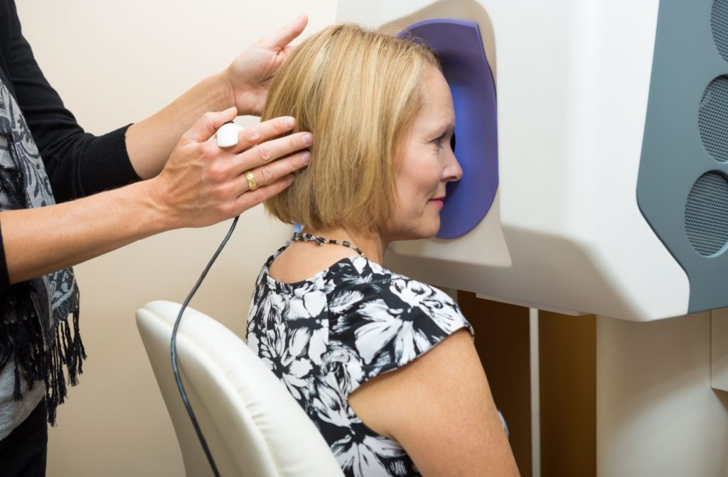
So, when you visit our team at Birring Eye Care, you might wonder: How do we check your retinas? This is done through a process called “retinal imaging.” This technology is used during eye exams to capture images of the back of your eye. The process is straightforward and often only takes a few seconds per eye.
To begin with, your optometrist may need to dilate your pupils with special eye drops, depending on their size. This lets more light enter the eye, making it significantly easier to see the retina.
Then, we use a noninvasive laser to scan your eye and send the images to a computer instantly. We can use these images to investigate your retina, catch any potential problems, and provide you with a diagnosis.
If we need to dilate your pupils, you’ll likely experience light sensitivity for a few hours after your exam. While it can help to wear a pair of sunglasses to reduce the sensitivity, you should avoid driving. Instead, contact a friend or family member to help you get home safely.

What to Expect During an Eye Exam
While retinal imaging is a crucial part of an eye exam, it isn’t the only test performed. We’ll quickly catch up on your family and medical history when you visit us for a comprehensive eye exam. This lets us determine if you’re at a higher risk of developing certain conditions. Then, we’ll likely check:
- Your visual acuity
- Your refraction abilities
- Your eye alignment
- The range of your visual field
- Your peripheral vision
- Your intraocular pressure
We may run additional tests to properly understand your eyes. Your vision is precious, and we like to be thorough!
Book Your Next Eye Exam
At Birring Eyecare, we take pride in our patient-first approach. We’re dedicated to serving our community for all their eye care needs and are here to help. Book your next appointment with our team, and let’s work together to protect your vision!